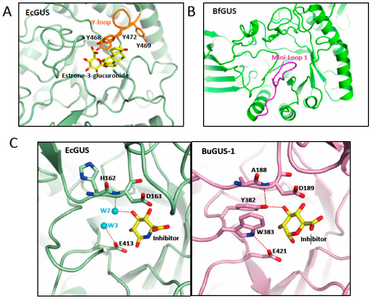
Figure 3.
Structural elements involved in the binding of glucuronides. Representation of the active center of GUS enzymes shown in (A) EcGUS (PDB 3K4D) (green) with the Y-loop and aromatic cage highlighted in orange and a molecule of estrone-3-glucuronide (yellow) modelled; (B) Mini-Loop 1 GUS of Bacteroides fragilis (PDB 3CMG) (green) highlighting the location of the mini-Loop 1 (magenta) involved in the binding of E-glucuronides. (C) Detail of the interactions of the water molecules 2 and 3 (cyan) in the active center of EcGUS (PDB 3K4D) (green) (left panel) compared with those observed in BuGUS-1 (pink) (right panel) involving residues Y382 and W383.

