Figure 3.
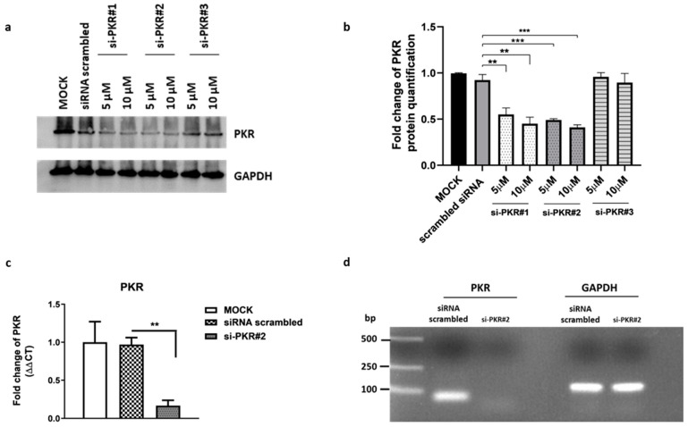
Validation of PKR silencing by western blotting and RT-qPCR. HEp-2 cells were transfected with three different siRNA duplexes at 5 and 10 µM to silencing PKR, and siRNA scrambled was used as negative control (10 µM). Samples were collected at 48 h post-transfection to measure the expression of PKR. (a) Western blot analysis was performed to detect the protein level of PKR following silencing. The GAPDH was used to check the protein loading. Panel (b) shows fold changes in protein quantification. Band density was determined with the Image J program, expressed as fold change over the appropriate housekeeping genes and with respect to siRNA scrambled control. The data were graphically represented by using GraphPad Prism 6 software. (c) Quantitative Real-time PCR was performed to evaluate the transcriptional levels of PKR in HEp-2 transfected with si-PKR#2 (10 µM) and siRNA scrambled. (d) The amount of PKR transcripts obtained following transfection with si-PKR#2 was confirmed by running the amplicons on an agarose gel. Statistical analyses were performed with one-way analysis of variance (ANOVA) in triplicate, and asterisks (** and ***) indicate the significance of p-values less than 0.01 and 0.001, respectively.

