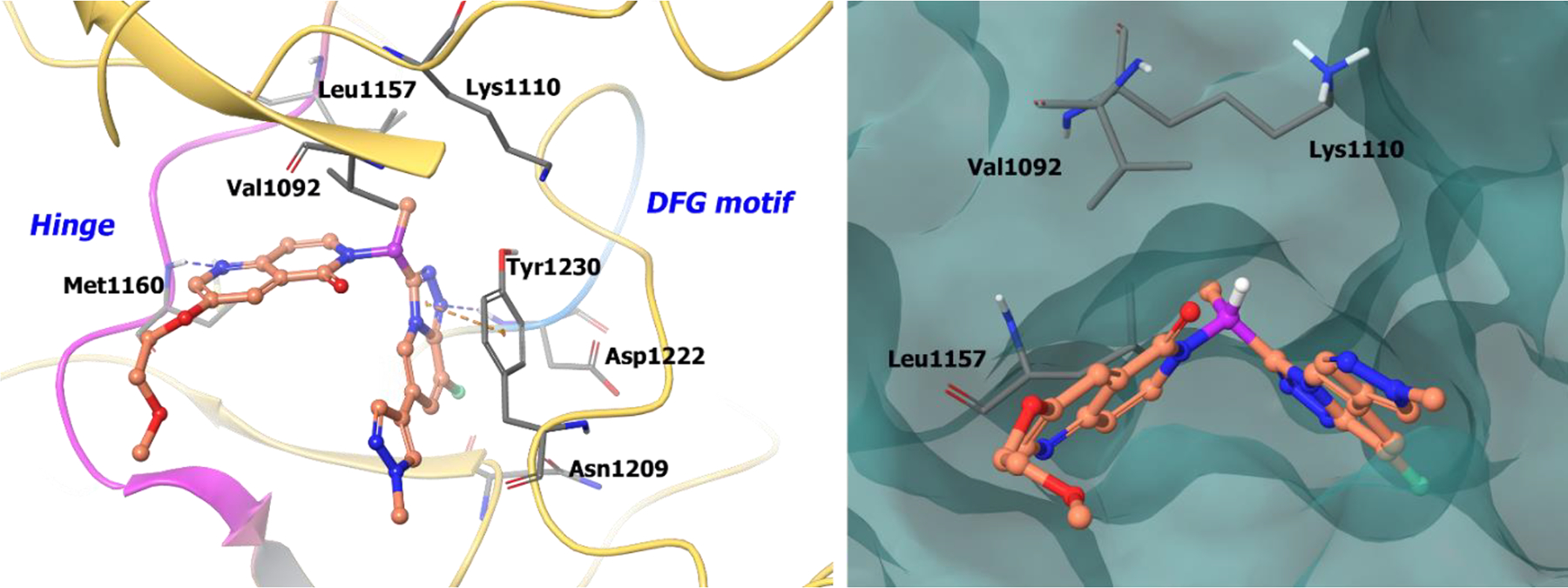
Figure 4.

The co-crystal structure of compound 16 bound to c-MET (PDB 5EYD, 1.85 Å). Left panel: Interaction of compound 16 with the protein. The protein is depicted as yellow ribbons, and the hydrogen bonds are illustrated with blue dashed lines; Right panel: Compound 16 binding site in c-MET, protein is shown as surface. Compound 16 atoms are colored as follows: carbon, red-orange; nitrogen, blue; oxygen, red; chlorine, green; fluorine, jade; hydrogen, white; the chiral carbon is highlighted in violet.
