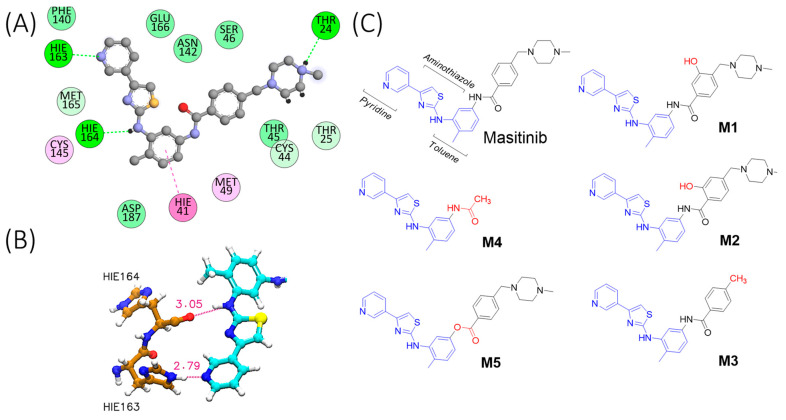
Figure 1.
Ligand–protein interactions and chemical structures. (A) Interactions between masitinib and Mpro residues. The small molecule is shown as grey chemical structure with heteroatoms highlighted in red (Oxygen), blue (Nitrogen) and yellow (Sulphur). The colours of the interactions correspond to π–π stacking (magenta), alkyl–π stacking (pink), hydrogen bonds (bright green), and Van der Waals interactions (green). The interaction maps were prepared using the BIOVIA Discovery Studio Visualizer [17]. (B) Schematic representation of key hydrogen bond interactions, displaying masitinib with His163 and His164. Hydrogen bonds are represented as dotted magenta lines, and the heavy distance (Å) is also shown with the same colour. The key residues His163 and His164 are shown in brown (Carbon), whereas masitinib (only a fragment, for simplicity) is depicted in cyan (Carbon). Heteroatoms are shown in red (Oxygen), blue (Nitrogen) and yellow (Sulphur). (C) Chemical structures of the proposed masitinib derivatives (M1–M5).

