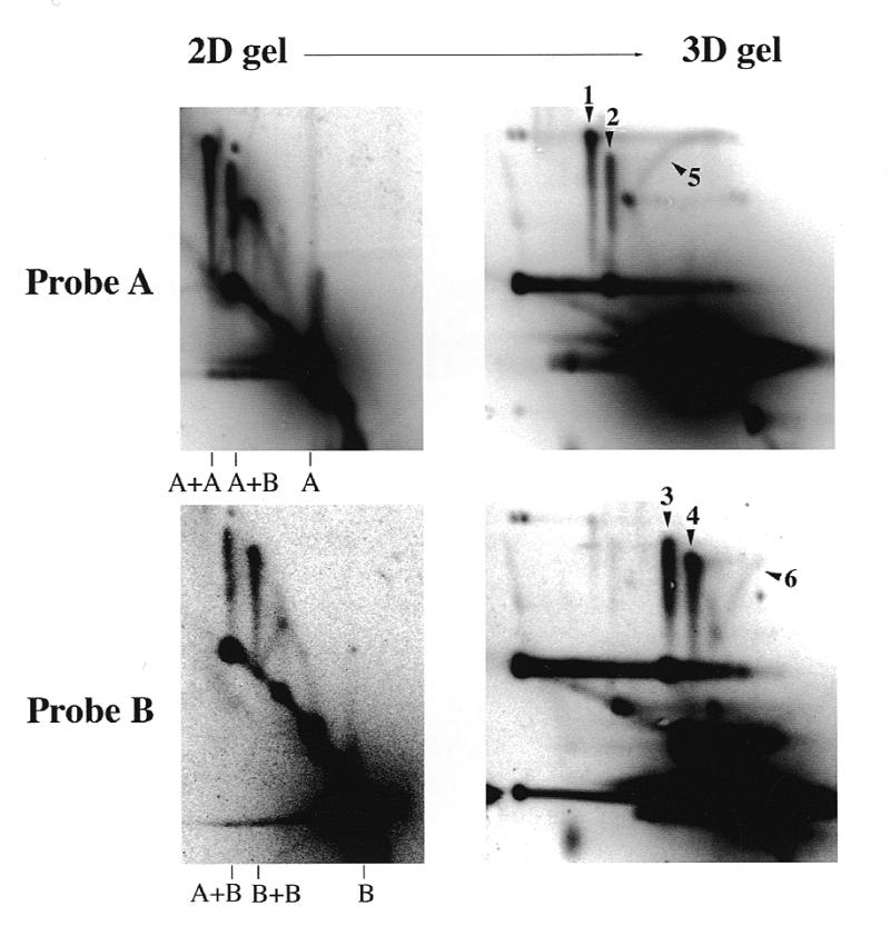
Figure 5.

2D (neutral–neutral) and 3D (neutral–neutral–alkaline) gel analysis of the junction component single DNA strands. pBR322 was incubated in Xenopus egg extracts for 90 min. DNA was purified and cut with AlwNI and BamHI and loaded on two duplicate 2D gels. One of the 2D gels was subjected to a third electrophoresis in alkali in the same direction as the first electrophoresis. After Southern blotting, the membranes were sequentially hybridised with the radioactively labelled A and B probes. Signals labelled 1–6 are described in the text.
