
Fig. 1.
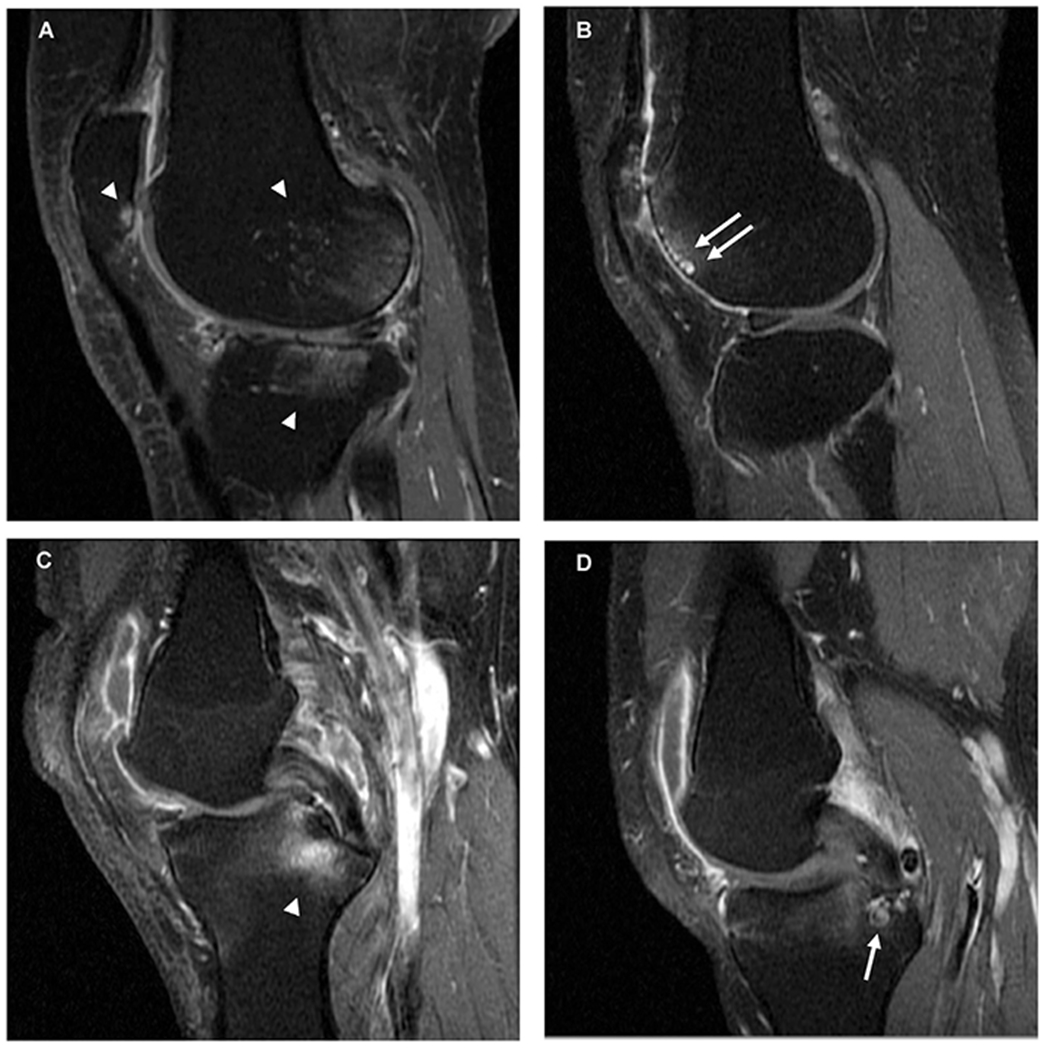
Bone marrow lesion (BML) types on sagittal T1-w fat suppressed contrast-enhanced MRI. (A) Subchondral BMLs at the posterior femoral condyle, posterior patella and lateral tibial plateau with ill-defined hyperintense (oedema-like) signal (white arrowheads). (B) Subchondral BML with cyst-like components (white arrows) in the anterior femoral condyle. (C) Ligament-based BML associated with the attachment of the posterior cruciate ligament (PCL) (odema-like signal extends into the subchondral bone) (white arrowhead). (D) Cyst-like component (white arrowhead) of a ligament-based BML in the tibia associated with the posterior cruciate ligament attachment.