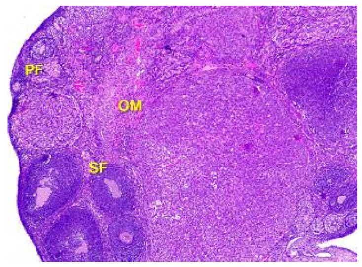
Figure 5.
Structural organization of the ovary cortex in an animal under the influence of a dose of 0.5 mg/kg of the body weight. Follicles in the cortex at different stages of the development: primary follicles (PF), secondary follicles (SF), Graafian follicles (GF). Blood vessels, elastic and collagen fibers in connective tissue stroma of ovarian medulla (OM). Stained with Hematoxylin and Eosin. ×40.

