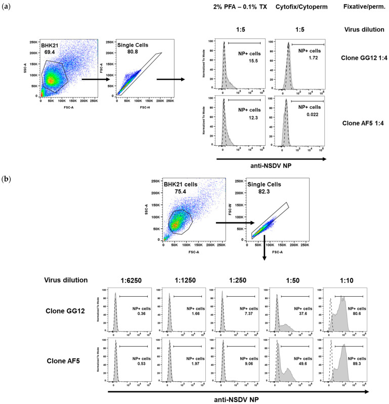
Figure 3.
Flow cytometry analysis of NSDV-infected BHK cells. (a) PFA/Triton X100 (PFA—0.1% TX) and Cytofix/Cytoperm treatments result in poor staining for both monoclonal antibodies used at a high concentration (1:4) and at a high concentration of virus (dilution of 1:5). (b) Methanol fixation-permeabilisation allows for staining of NSDV-infected cells in a dose-dependent manner. Cells were infected with varying dilutions of virus (1:10 to 1:6250) and both antibodies were used at 1:50. For (a,b), the gating strategy is provided; dashed line in histograms represent NP staining of uninfected BHK cells while grey curve represents NP staining for BHK-infected cells.

