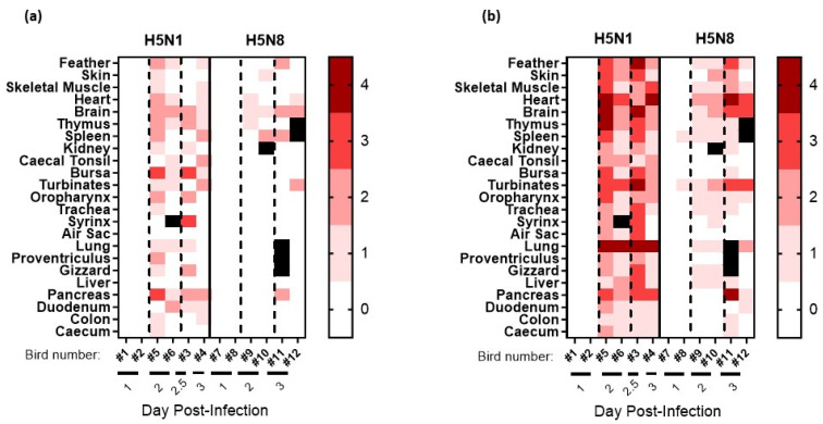
Figure 4.
Semi-quantitative scoring of (a) histopathological changes visualised via H&E staining and (b) tissue distribution of viral-specific NP antigen determined with IHC from chickens directly infected with high-dose H5N8-2020 or H5N1-2020 during the pathogenesis time-course investigation. Tissue samples were collected at 1, 2, and 3 dpi. Viral-specific antigen staining was scored according to the following scale: 0 absent; 1 rare; 2 scattered; 3 confluent; and 4 abundant. Black squares indicate that no IHC scoring was carried out.

