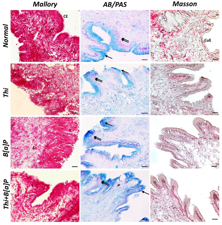
Figure 1.
Cross section of mantle of M. galloprovincialis (40×, scale bar: 10 µm). Mallory trichrome and Masson trichrome morphological staining revealed a homogeneous, well-organized connective tissue (C) and an epithelium with columnar (CE) and cuboidal (CuE) cells in control animals. AB/PAS histochemical staining revealed the presence of acidic (arrow) and neutral (double arrow) mucopolysaccharides. In groups exposed to pollutants, the epithelium was more disorganized, with looser connective tissue and an overproduction of acidic mucopolysaccharides.

