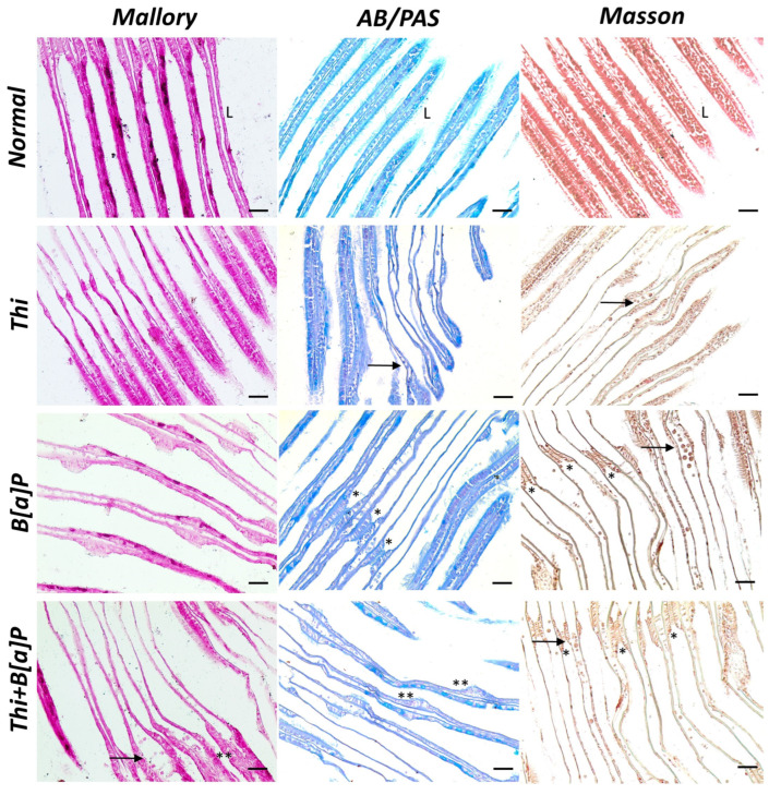
Figure 2.
Cross section of gills of M. galloprovincialis (40×, scale bar: 10 µm). Control specimens showed well-organized, linear gills with homogeneous lamellae (L) and compact epithelium. In the exposed groups, the lamellae appear disorganized, and the epithelial tissue appear inhomogeneous. Fusion of lamellae (*) with hyperplastic and hypertrophic cells (**) is also evident in some places. In addition, aggregates of hemocytes are evident in the pollutant-exposed groups (arrow).

