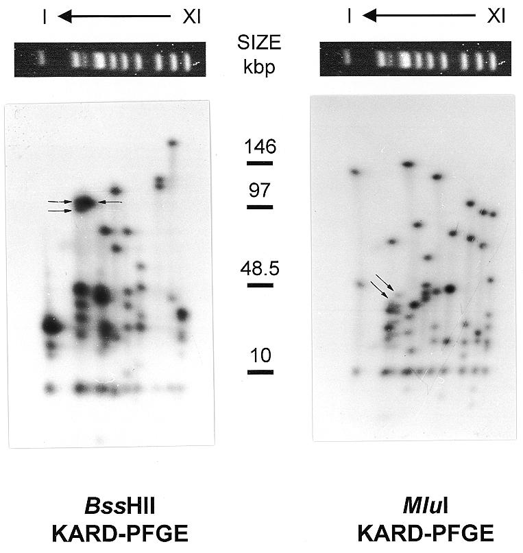
Figure 1.

KARD-PFGE of the E.cuniculi genome. Autoradiographs of radiolabelled chromosomal DNA are shown after resolution of the molecular karyotype (at the top of the 2D-gel) and in-gel digestion with BssHII (left part) or MluI (right part) followed by a second orthogonal PFGE. Chromosome order is indicated above, the size of standards (in kb) in the middle. The two restriction fragments derived from chromosomes IIIa and IIIb, which can be differentiated by their size, are indicated by arrows. The decreasing intensity of spots in the right part of the lower region of BssHII KARD autoradiograph is due to a heterogeneous drying of the gel.
