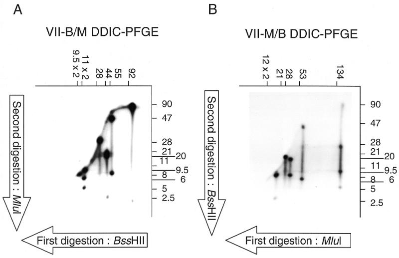
Figure 2.

DDIC-PFGE of the chromosome VII of E.cuniculi. After isolation of the chromosome VII from the molecular karyotype, chromosomal DNA was digested by BssHII (A) or MluI (B), and radiolabelled. A second PFGE was performed leading to the separation of fragments (sizes in kb, above the autoradiograph). A second digestion was then performed with MluI (A) or BssHII (B), and the fragments separated with a third PFGE, orthogonal to the second (sizes in kb, on the right of each autoradiograph). Autoradiographs on (A) and (B) are therefore called VII B/M DDIC-PFGE and VII M/B DDIC-PFGE, respectively.
