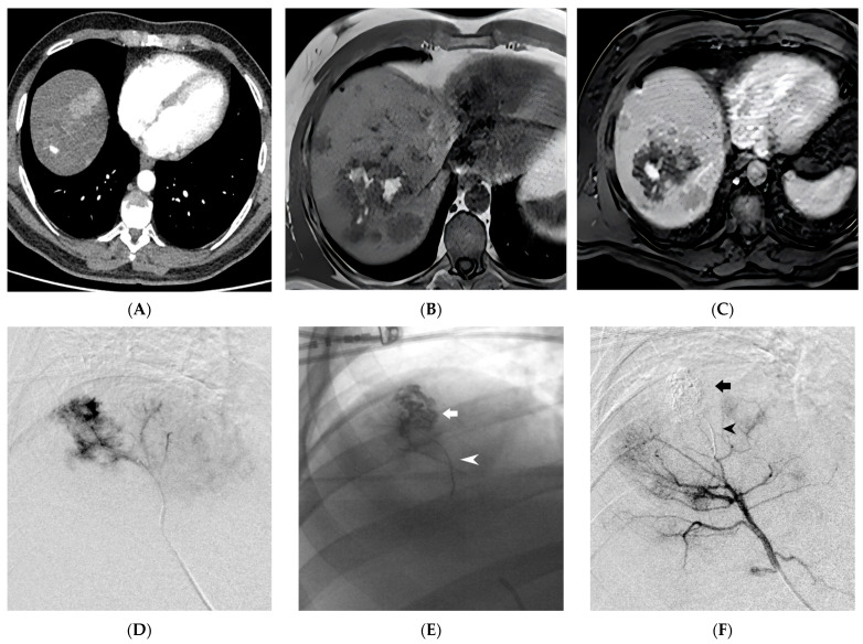
Figure 3.
Ruptured Hepatocellular Carcinoma (HCC) within the hepatic parenchyma is depicted in the images. In the contrast-enhanced Computed Tomography, arterial phase, there is evidence of a 1 cm pseudoaneurysm within an HCC nodule (A). On the Magnetic Resonance Imaging (MRI) T1 Fast Field Echo In-Phase sequence, hyperintense material (blood) is observed within a large HCC nodule of the right lobe (B). Gadobenate dimeglumine (Gd-BOPTA)-enhanced MRI in the arterial phase confirms the presence of an intranodular contrast blush (C). Superselective digital subtraction angiography of the S7 hepatic artery branch shows contrast leakage into the ruptured HCC (D). Successful embolization of the HCC nodule (indicated by the arrow) and the parent artery (indicated by the arrowhead) was achieved using Ethylene-Vinyl Alcohol (EVOH) copolymer (E,F). (From Minici et al. doi: 10.3390/medicina59040710, by MDPI, Basel, Switzerland, licensed under CC BY 4.0).

