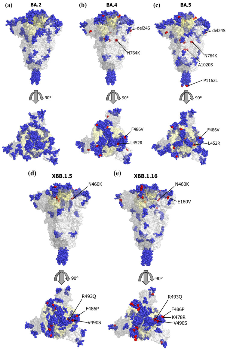
Figure 4.
Conformational epitopes and main mutations mapped on three-dimensional structure models of SARS-CoV-2 S protein. We mapped the predicted conformational epitopes on three-dimensional structure models of SARS-CoV-2 S protein, including the receptor-binding domain (RBD). The chains of the trimer structures are colored in dark gray (chain A), gray (chain B), and light gray (chain C). Conformational epitopes, main mutation sites, and RBD are shown in blue, red, and light-yellow colors, respectively. (a) SARS-CoV-2 BA.2 variant S protein 3D structure; (b) SARS-CoV-2 BA.4 variant S protein 3D structure; (c) SARS-CoV-2 BA.5 variant S protein 3D structure; (d) SARS-CoV-2 XBB.1.5 variant S protein 3D structure; (e) SARS-CoV-2 XBB.1.16 variant S protein 3D structure.

