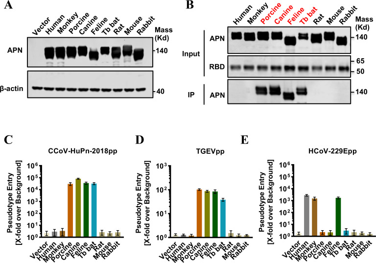
FIG 2.
Multiple APN orthologs served as receptors for CCoV-HuPn-2018. (A) Transient expression of APN orthologs in 293T cells. The cell lysates were detected by western blot assay using an anti-C9 monoclonal antibody. β-Actin served as a loading control. (B) IP assay. The upper panel shows the input of APN protein with a C9 tag and RBD with an IgG Fc tag (RBD-Ig). The lower panel shows the APN pulled down by the RBD-Ig fusion protein. (C–E) Pseudotyped virus entry of CCoV-HuPn-2018 (C), TGEV (D), and HCoV-229E (E). 293T cells were transfected with the empty vector pcDNA3.1 or APN orthologs. At 48 h post-transfection, the cells were infected by the pseudotyped viruses CCoV-HuPn-2018 (CCoV-HuPn-2018pp), TGEV (TGEVpp), and TGEV (TGEVpp). At 24 h post-infection, pseudotype entry was determined by measuring luciferase activity in cell lysates. Luciferase signals obtained for particles bearing no envelope protein were used for normalization. Error bars reveal the standard deviation of the means from three biological repeats.

