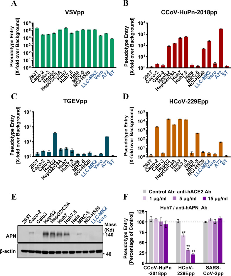
FIG 4.
Entry of CCoV-HuPn-2018 into multiple human cell lines in a hAPN-independent manner. (A to D) Cell lines of human and other animal origins were infected with VSV-Gpp (A), CCoV-HuPn-2018pp (B), TGEV (C), or HCoV-229Epp (D). At 24 h post-infection, luciferase signals were measured, and pseudotype entry was normalized against the background (a luciferase signal was obtained for pseudotyped particles without viral envelope protein). Error bars reveal the standard deviation of the means from three biological repeats. Four animal-derived cell lines are shown in dark blue. (E) The levels of endogenous APN proteins were determined by Western blot assay using an anti-APN polyclonal antibody, and β-actin served as a loading control. (F) Entry inhibition assay by anti-hAPN antibody. Huh7 cells were pre-incubated with the indicated concentration of anti-hAPN antibody (or anti-hACE2 antibody as a control) for 1 h and then infected with CCoV-HuPn-2018pp in the presence of the indicated concentration of hACE2 antibody or hAPN antibody for another 3 h, and then the virus and antibodies were removed and replaced with fresh medium for further incubation. At 24 h post-infection, luciferase signals were measured, and pseudotype entry was normalized against the control antibody-treated cells. Error bars reveal the standard deviation of the means from three biological repeats. **, P < 0.001 compared to the level of the control antibody.

