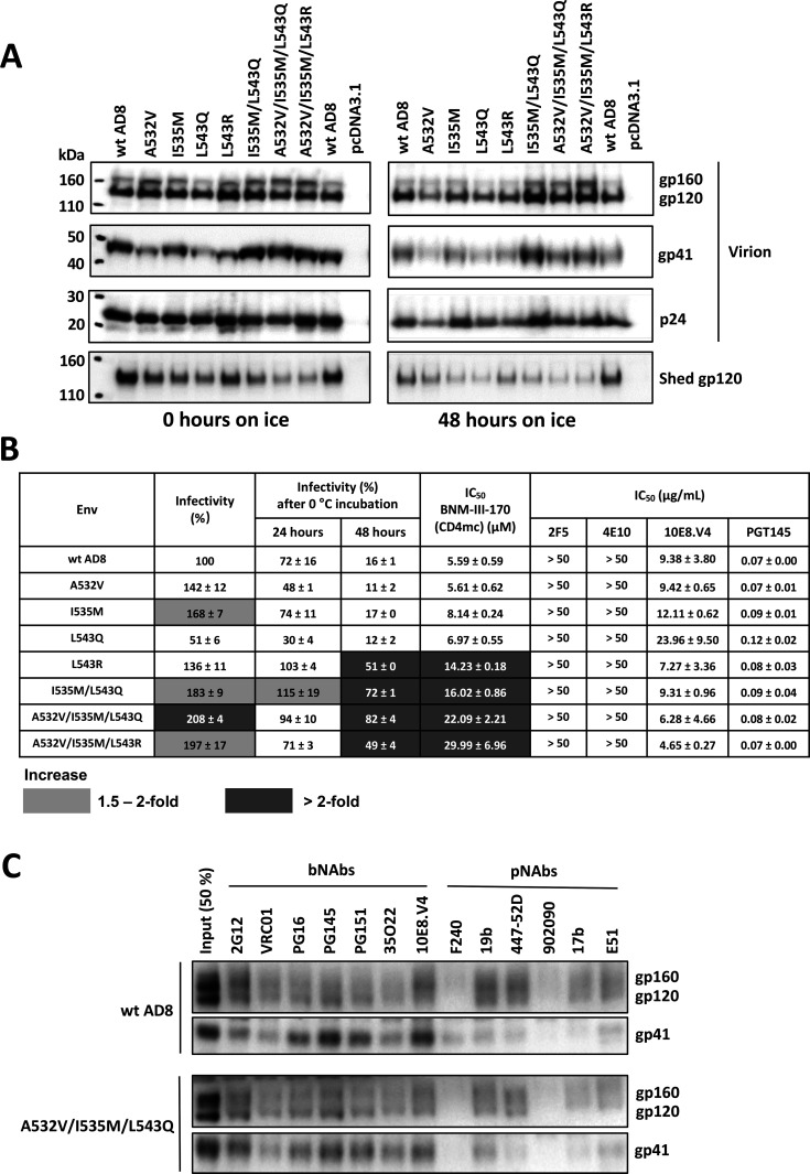
Fig 10.
Analysis of HIV-1AD8 Env FPPR mutants expressed from an infectious HIV-1 proviral clone. (A) HEK 293T cells were transfected with plasmids containing an infectious NL4-3 provirus with the envs expressing the indicated wild-type (wt) or mutant AD8 Bam Envs. The Bam changes (S752F I756F) restore the natural HIV-1AD8 Env sequence near the AD8/HXBc2 junction and reduce the clipping of the gp41 cytoplasmic tail by the viral protease (90). Seventy-two hours later, the cell supernatants were collected, filtered (0.45 µm), and aliquoted. The virus-containing supernatants were incubated on ice for 0 or 48 h, after which they were centrifuged at 14,000 × g for 1 h at 4°C. The pellets were resuspended in 1× PBS and represent the virion samples. The supernatants were incubated with GNL beads for 1.5 h at room temperature; the GNL precipitates represent the shed gp120. Virion and shed gp120 samples were western blotted with a goat anti-gp120 antibody. (B) The infectivity of the virions with the indicated HIV-1AD8 Env variants without treatment or after 24 and 48 h of incubation on ice was measured on TZM-bl cells. The values are normalized to that of the untreated wt AD8 Bam virions. The fold increases observed for the Env mutants relative to the values for the wt AD8 Bam virus under the same treatment conditions are colored according to the key. The inhibition of virus infectivity after 1 h incubation at 37°C with the CD4mc, BNM-III-170, or antibodies is reported. The 50% inhibitory concentrations (IC50 values) are reported in µM for BNM-III-170 and in µg/mL for the antibodies. Note that all the viruses were resistant to the 2F5 and 4E10 bNAbs against the gp41 membrane-proximal external region (MPER). The results shown are the means and SDs derived from two independent experiments. Inhibition by BNM-III-170 is colored according to the key. (C) Purified virus particles were incubated with a panel of broadly neutralizing antibodies (bNAbs) and poorly neutralizing antibodies (pNAbs) for 1 h at room temperature. The virus-antibody mixture was washed with 1× PBS and centrifuged. The virus-antibody pellet was lysed and precipitated with Protein A-agarose beads for 1 h at 4°C. The beads were washed three times and western blotted with a goat anti-gp120 antibody and the 4E10 anti-gp41 antibody.

