Figure 6.
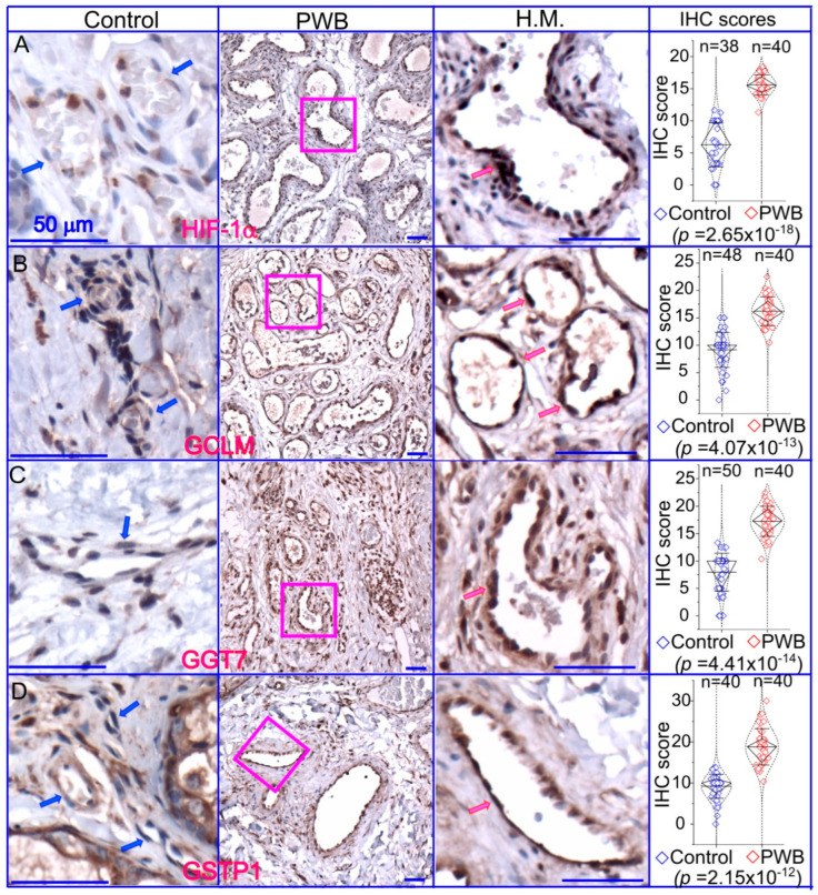
Expressions of key molecules associated with glutathione metabolism in PWB lesions. The IHC assays using antibodies against HIF−1α (A), GCLM (B), GGT7 (C), and GSTP1 (D) show the immunoreactive blood vessels in control skin or PWB lesions. H.M., a higher magnification from the pink boxed area in the left panel showing immunoreactive positive PWB blood vessels for the corresponding antibodies. Scale bar: 50 µm. n, number of blood vessels from PWB (4 subjects) or normal ones (5 subjects). Whiskers: mean ± S.D.; Diamond boxes: IQR; Dotted curves: data distribution. Paired t-test was used for comparing arbitary IHC scores. Blue arrows: control dermal capillaries; red arrows: PWB blood vessels.

