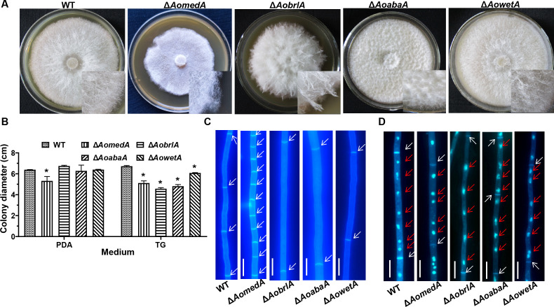
Fig 4.
Comparison of mycelial growth, septa, and nuclei between WT and mutant strains. (A) Colony morphology of WT and mutant strains incubated on TYGA medium for 5 d. (B) Comparison of colony diameters of WT and mutant strains incubated on PDA and TG medium for 5 d. Error bars: SD from three replicates. Asterisk indicates a significant difference between mutant and WT strain (Tukey’s HSD, P < 0.05). (C) Mycelial cells were stained with CFW dye. White arrows indicate cell septa of the hyphae. (D) The nuclei of mycelial cells were stained using CFW and DAPI. White arrows indicate the cell septa of hyphae, and red arrows indicate nuclei. Bar = 10 µm.

