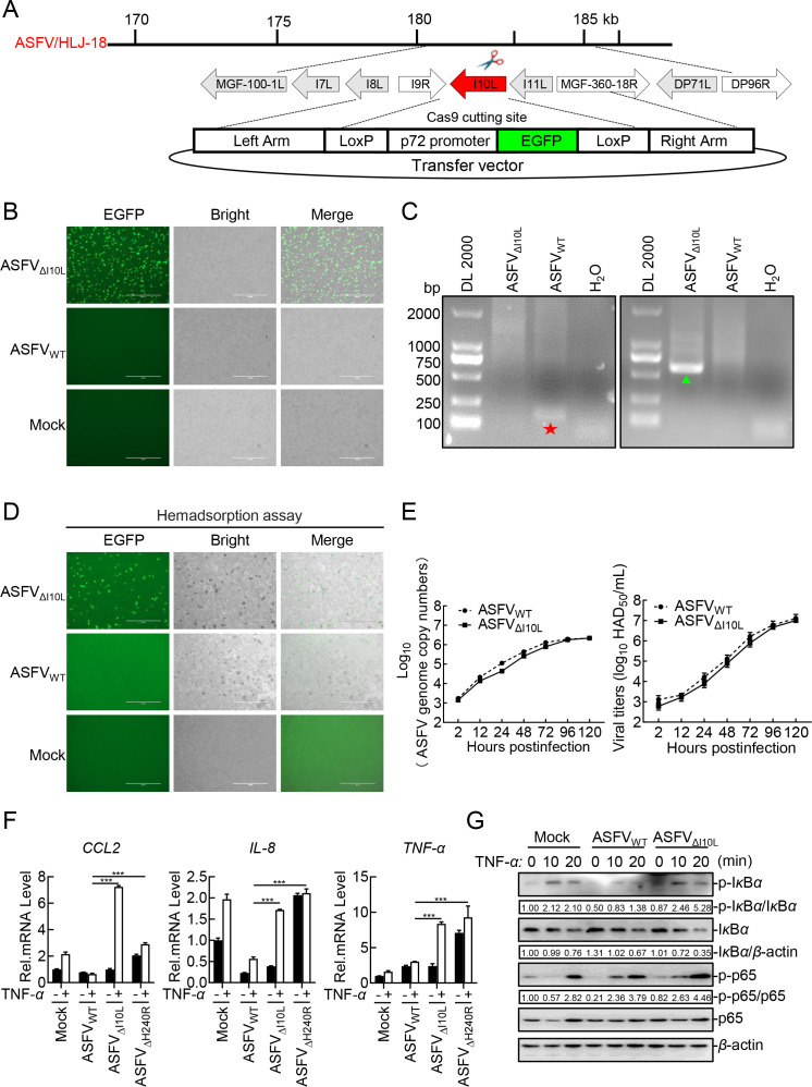
Fig 3.
Deletion of the I10L gene from the ASFV genome results in enhanced activation of the NF-κB signaling pathway. (A) Schematic diagram of the genome structure of ASFVΔI10L. (B) Purification of ASFVΔI10L in primary porcine alveolar macrophages (PAMs). The recombinant ASFVΔI10L was screened out relying on EGFP fluorescence detection. (C) Identification of ASFVΔI10L by PCR. Specific primers of I10L (★) and EGFP (▲) were used for PCR, and the samples were subjected to agarose gel electrophoresis. (D) Hemadsorption characteristics of ASFVΔI10L. PAMs were infected with ASFVΔI10L or ASFVWT or left uninfected serving as a negative control. Hemadsorption and fluorescence were examined by microscopy. (E) In vitro replication characteristics of ASFVΔI10L. Left panel: genome copies of ASFVΔI10L or ASFVWT; Right panel: viral titers of ASFVΔI10L or ASFVWT. (F) ASFVΔI10L induces much higher levels of CCL2, IL-8, and TNF-α than did ASFVWT. PAMs were either mock-infected or infected with ASFVWT, ASFVΔI10L or ASFVΔH240R at a multiplication of infection (MOI) of one. Forty-eight hours later, the cells were treated with TNF-α (10 ng/mL) (or left untreated) for another 20 minutes. Total RNA was extracted and subjected to RT-qPCR. (G) ASFVΔI10L elevates the phosphorylation levels of IκBα and p65. PAMs were left uninfected or infected with ASFVWT or ASFVΔI10L (MOI = 3) for 24 hours. The cells were then treated or untreated with TNF-α (20 ng/mL) for 10 or 20 minutes, immunoblotting analysis was performed. Densitometric analysis of protein expression levels were performed using ImageJ software. Data shown are the mean ± SD from one representative experiment performed in triplicates. ***P < 0.001 (unpaired t test).

