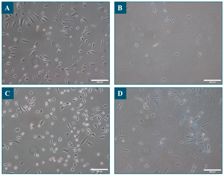
Figure 4.
Bright-field microscopy of sebocytes grown with or without bacteria. Images were taken 48 h after the inoculation of the bacteria, just after removing the culture supernatant containing the bacteria and just before performing the Alamar Blue test. Panel (A): sebocytes only; Panel (B): sebocytes and S. epidermidis; Panel (C): sebocytes and C. acnes; Panel (D): sebocytes and C. acnes and S. epidermidis. Scale bar: 200 µm.

