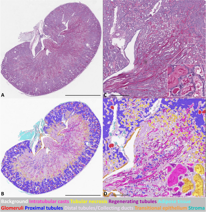
Fig. 1.
CNN-based automated segmentation on WSI of an injured murine kidney on day 3 after IRI. (A) PAS stained WSI; (B) corresponding segmentation result of WSI in A. (C) High-magnification PAS stained WSI, with further higher magnification area (inset); (D) corresponding segmentation result of WSI in C, with further higher magnification area (inset). Scale bars: 2 mm (A,B); 400 um (micrometers) (C,D).

