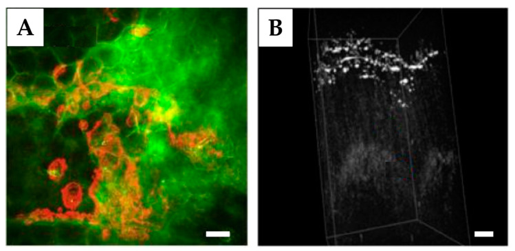
Figure 8.
Representative SRS (A) and OCT (B) images of skin penetration. (A): Deposition of the ruxolitinib (red) in the SC of mouse skin (green) ex vivo measured with SRS (“Stokes” beam at 803 nm to target lipids at 2845 cm−1 and “pump” beam at 845 nm to target the nitrile stretch of ruxolitinib at 2220 cm−1), reprinted with permission from Ref. [279]; (B): Penetration of nanodiamonds into mice skin ex vivo (white areas with strong scattering show the presence of nanodiamond clusters in the epidermis) measured with OCT (excitation at 930 nm), adopted under CC BY 4.0 license from Ref. [102]. Scale bar: 20 µm (A), 150 µm (B).

