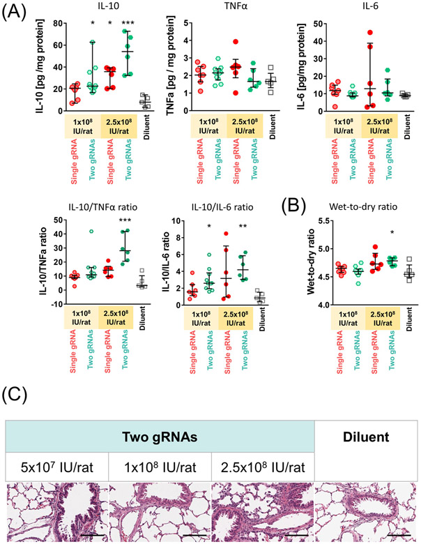
Figure 5.
Analysis of protein levels and histology of the lungs treated with adenoviral-vector mediated IL-10 activation. (A) Concentrations of each target protein in the lung tissue were measured by enzyme-linked immunosorbent assay (ELISA) using lung tissues collected 3 days after the treatment (1x10⁸ IU/rat single gRNA, n=7; 1x10⁸ IU/rat two gRNAs, n=10; 2.5x10⁸ IU/rat single gRNA and two gRNAs, n=6; and Diluent, n=5). IL-10/TNF-α and IL-10/IL-6 ratio were calculated using the concentrations of each protein obtained by ELISA. (B) Wet-to-dry weight ratios of the lung tissues after 3 days of the vector administration. Median (interquartile range) in the same cases as in A are shown. A Kruskal-Wallis test followed by Dunn’s correction was used for comparison. Symbols indicate: *: p ≤ 0.05, **: p ≤ 0.01, ***: p ≤ 0.001. (C) Representative images of hematoxylin and eosin staining of the lung tissues fixed at day 3. Scale bars, 100 μm.

