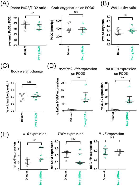
Figure 8.
Assessment of IL-10-activated donor lungs in syngeneic single lung transplant non-injury survival model. Two-gRNA adenoviral vector or buffer only (diluent group) was delivered to left lung of donor Lewis rats in vivo using the same immunosuppression protocol illustrated in Fig. 3A. After 24h of viral delivery, the left lung grafts were transplanted into recipient Lewis rats. Grafts were assessed on postoperative day 3 (POD3). Recipients received triple immunosuppression until analysis (n=5 and n=6 for diluent and two gRNA group, respectively). (A) Donor PaO2/FiO2 ratio calculated from PaO2 of systemic arterial blood taken at FiO2 of 0.5 (left). Graft oxygenation on POD0 was assessed by measuring PaO2 of blood taken from the left pulmonary vein after reperfusion (right). (B) Transgene and rat IL-10 expression in the left lung graft on POD3 measured by qPCR. Values from the left lungs of untreated Lewis rats (n=2) were set as 1. (C) Lung tissue of grafts collected on POD3 were stained for SaCas9 (green) and rat IL-10 mRNA (red) using fluorescence in situ hybridization. Nuclei were counterstained with DAPI. The scale bar indicates 20 μm. (D) Wet-to-dry ratio of left lung graft collected on POD3. (E) Representative inflammatory gene expression was measured by qPCR. Values from the left lungs of untreated Lewis rats (n=2) were set as 1. (F) Recipient body weight on POD3 shown as a percentage of those on POD0. Median(interquartile range) is shown. The Mann-Whitney test was used for statistical comparisons. NS: not significant, **: p ≤ 0.01.

