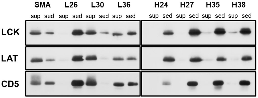
Figure 2.

SDS PAGE/Western blot analysis of Jurkat T-cell membranes solubilized by the SSS copolymers. The cell membranes were treated with the lysis buffer containing 1% of a copolymer, and the insoluble material was removed by centrifugation. Three typical membrane proteins (CD5, LCK, LAT) were detected by immunostaining in the sediments and supernatants of the copolymer-treated membrane samples. Only the relevant parts of the blots are shown, corresponding to the area around the MW of the respective proteins (55 kDa for LCK, 45 kDa for LAT and 65 kDa for CD5). Sed – sediment, sup – supernatant.
