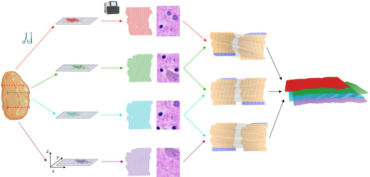
Figure 1.
PASTE2 partial alignment of overlapping slices. Four thin slices (red, green, blue, purple) are dissected from the same tissue and placed on an ST array. However, these slices only partially overlap in the z-coordinate direction. The inputs to PASTE2 are the four ST slices, including gene expression, spot locations, and optionally, histology images. PASTE2 computes a partial alignment of each pair of adjacent slices by selecting subsets of spots from each slice that preserve transcriptional, spatial, and image similarity. PASTE2 uses the partial alignment to create a 3D spatial reconstruction of the tissue.

