
Figure 3.
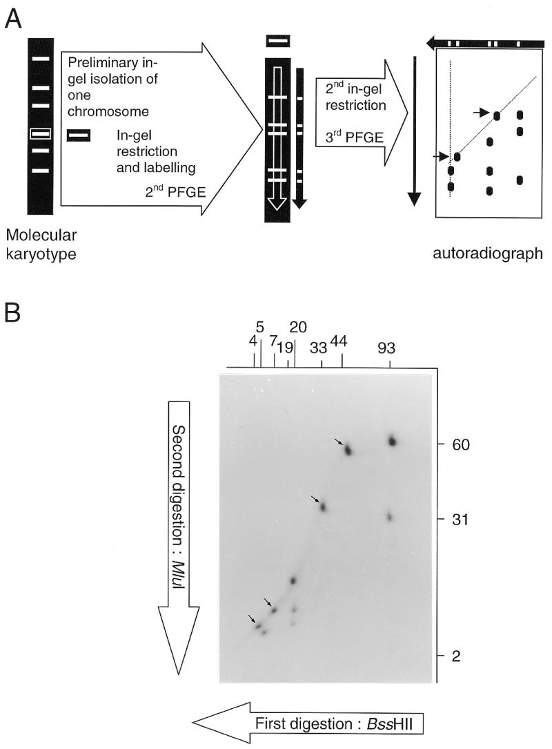
DDIC-PFGE. (A) Principle of the procedure. Separated DNA fragments arise from two successive in-gel restrictions of an individual chromosome. (B) Autoradiograph obtained after BssHII–MluI DDIC-PFGE of the chromosome I of S.cerevisiae YPH80. The first enzyme (BssHII) gives fragments (sizes indicated in kb above the autoradiograph) as determined by KARD-PFGE (see Fig. 2). The second enzyme (MluI) generates new fragments of which the sizes are indicated in kb at the right of the autoradiograph. Arrows show spots corresponding to fragments devoid of any restriction site for the second enzyme.