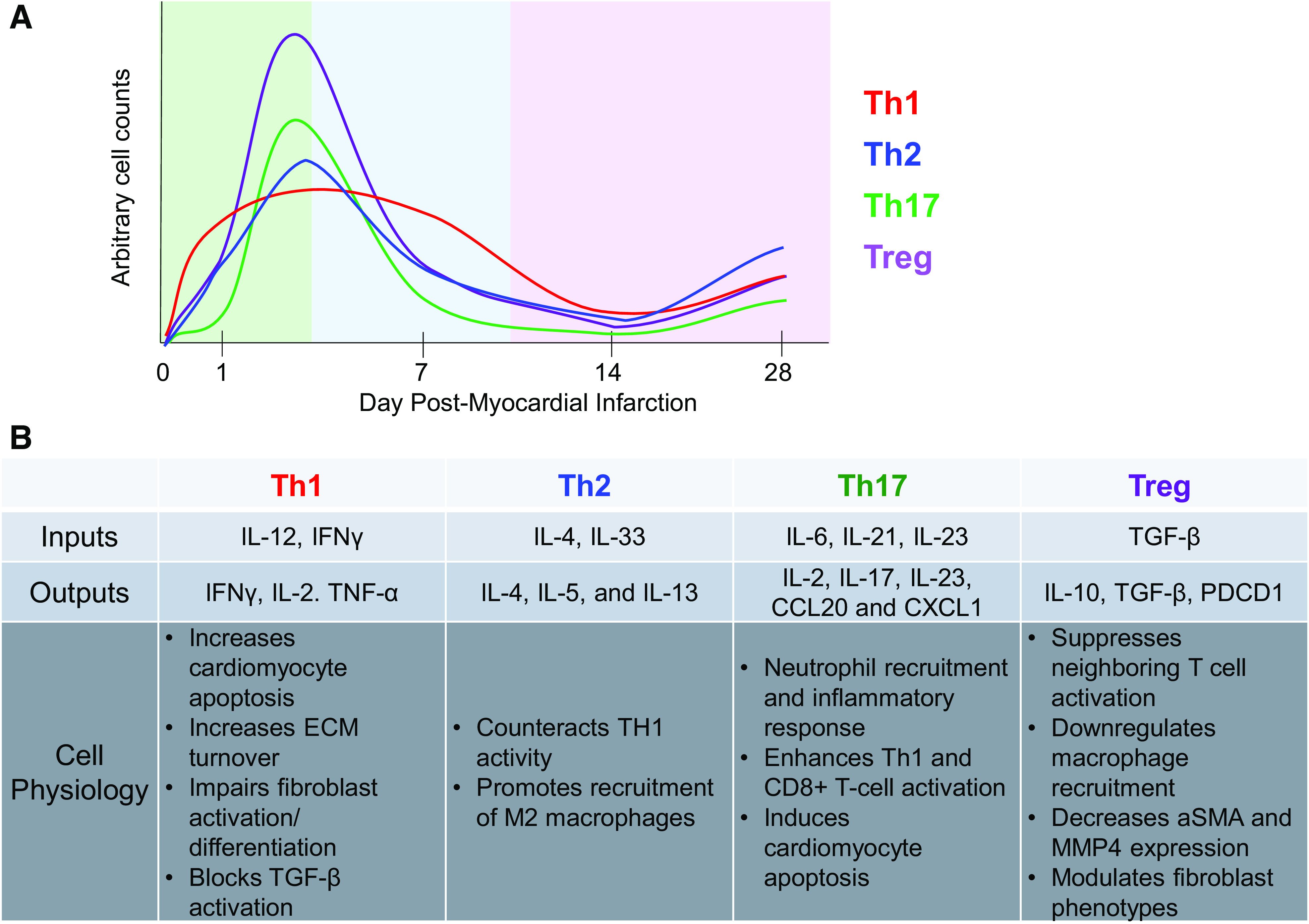
Figure 2.

A: temporal phenotypic changes in CD4+ T cells during the inflammation (green), proliferation (blue), and maturation (purple) phases of cardiac remodeling post-MI. Arbitrary cell counts mapped are based on findings presented in Kumar et al. (25). B: summary of the roles of CD4+ T cells postmyocardial infarction in the permanent occlusion model. α-SMA, α-smooth muscle actin; ECM, extracellular matrix; IFN, interferon; IL, interleukin; MMP, matrix metalloproteinase; PDCD, programmed cell death protein; TNFα, tumor necrosis factor α; TGF-β, tissue growth factor-β.
