
Figure 5. Distribution of Ell in the mid-ventricle.
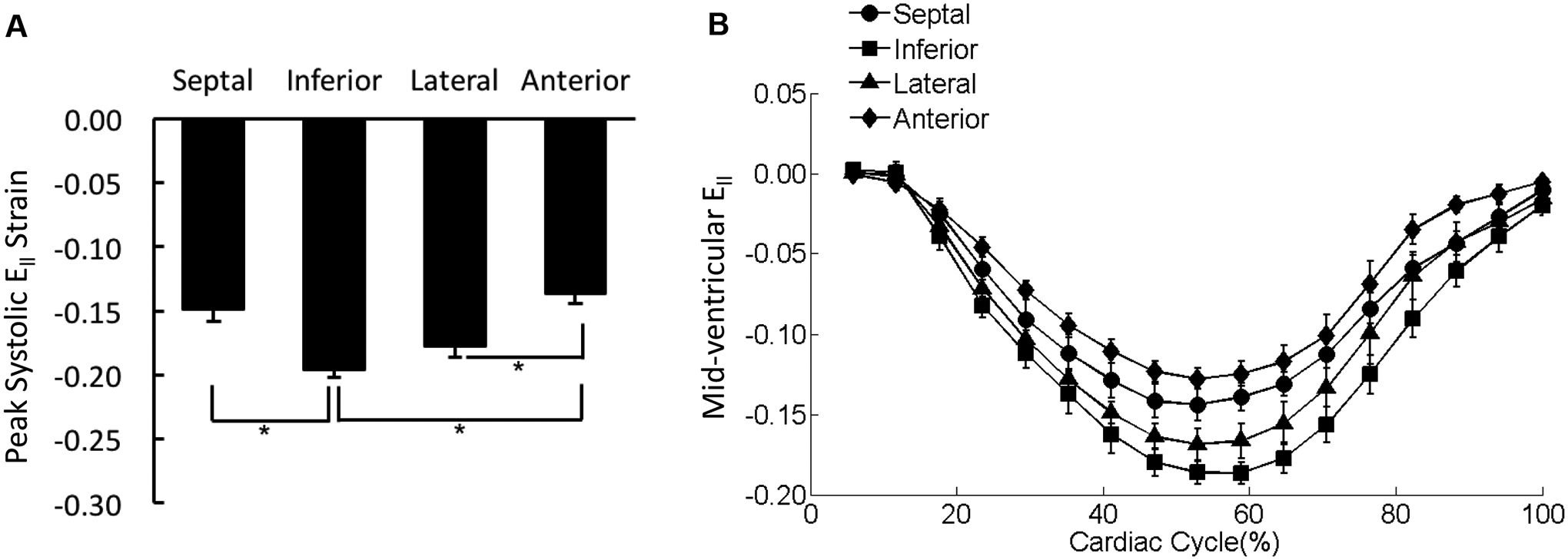
(A) The peak systolic Ell values were averaged over each of the four wall segments of the mid-ventricular slice regardless the transmural location. *p < 0.05. (B) The strain-time curves for Ell in the four wall segments of mid-ventricular slice were overlaid. Cardiac cycle started with end diastolic state.