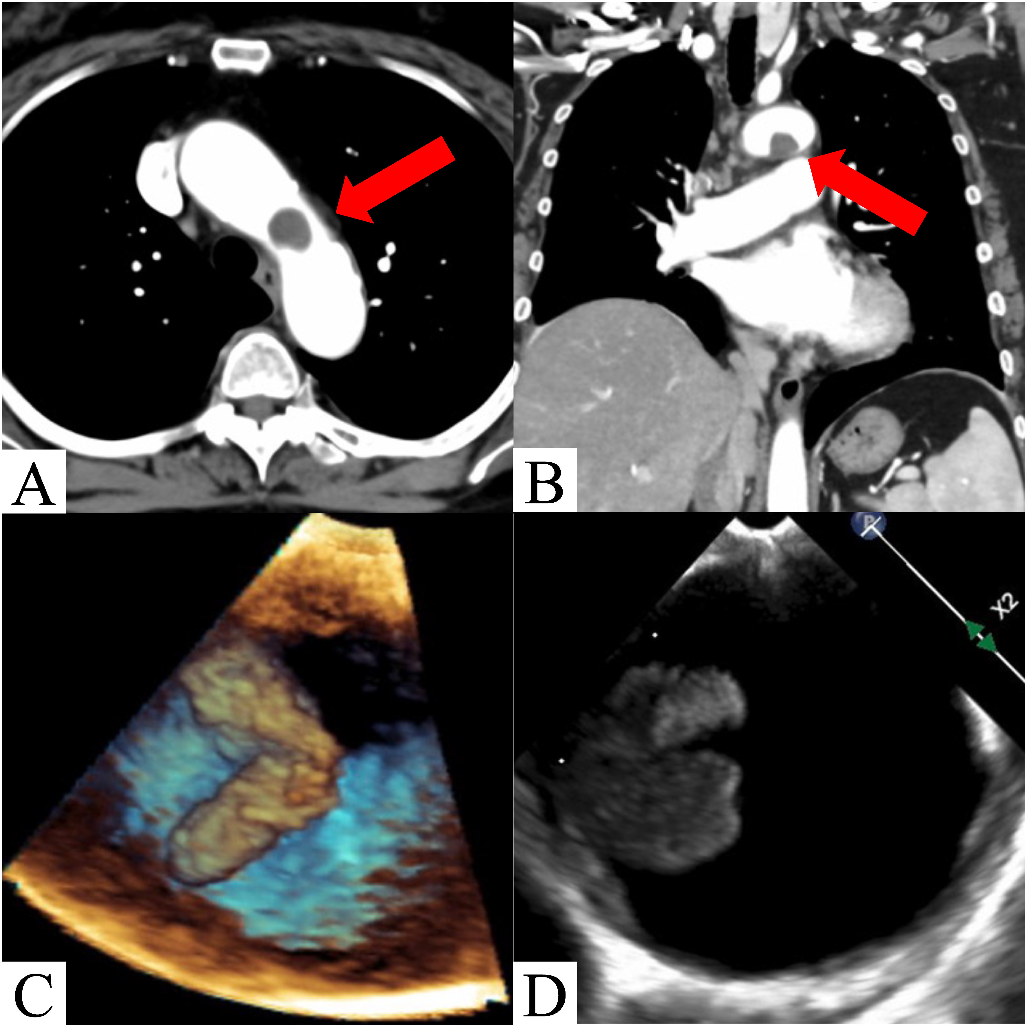
Fig. 1 Contrast-enhanced computed tomography (CT) and transesophageal echocardiography image findings. (A, B) Preoperative contrast-enhanced CT scan shows a 16×12 mm mass in the aortic arch. (C, D) Transesophageal echocardiography shows an approximately 15 mm mobile mass in the aortic arch.