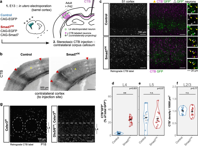
Fig. 4. Output connectivity of Smad7OE neurons.
a Schematic illustrating stereotaxic CTB injections into the corpus callosum to label contralaterally-projecting neurons in barrel cortex of adult control and Smad7OE brains. b Retrogradely labeled (CTB+) neurons in the cortex contralateral to the injected corpus callosum. A high degree of retrograde label is found in supra- and infra-granular layers in both genotypes. In adults, L4 is sparsely labeled in controls compared to Smad7OE (yellow asterisk). Anterograde labeling can also be observed as diffuse axonal signals (red arrowheads). c Retrogradely-labeled (CTB+) transfected L4 (GFP+) neurons in barrel cortex, contralateral to the injected area. Within L4, sparsely-distributed CTB+ neurons were labeled in controls. With Smad7OE, an increased amount of CTB labeling was observed throughout L4. Example regions showing double-positive CTB+/GFP+ (yellow triangles) and CTB-GFP+ (open triangles) transfected neurons are shown on the right. d–f Quantification of CTB+ neurons displayed as percentage of double-positive CTB+/GFP+ (of total number of electroporated GFP+ neurons) in L4 (d), L5 (e), and CTB+ density in L2/3 (f). Violin plots show distributions from 6 sections (on average) from 3 brains per genotype. Group means are displayed with error bars representing SEM. Asterisks indicate **p = 0.001, linear mixed-effects model with parametric bootstrap test (n = 19 sections from 3 control brains, n = 20 sections from 3 Smad7OE brains). g Labeling of retrogradely projecting neurons in somatosensory cortex of Celsr3 control (flox/flox) and cKO brains at P18. Source data are provided as a Source data file.

