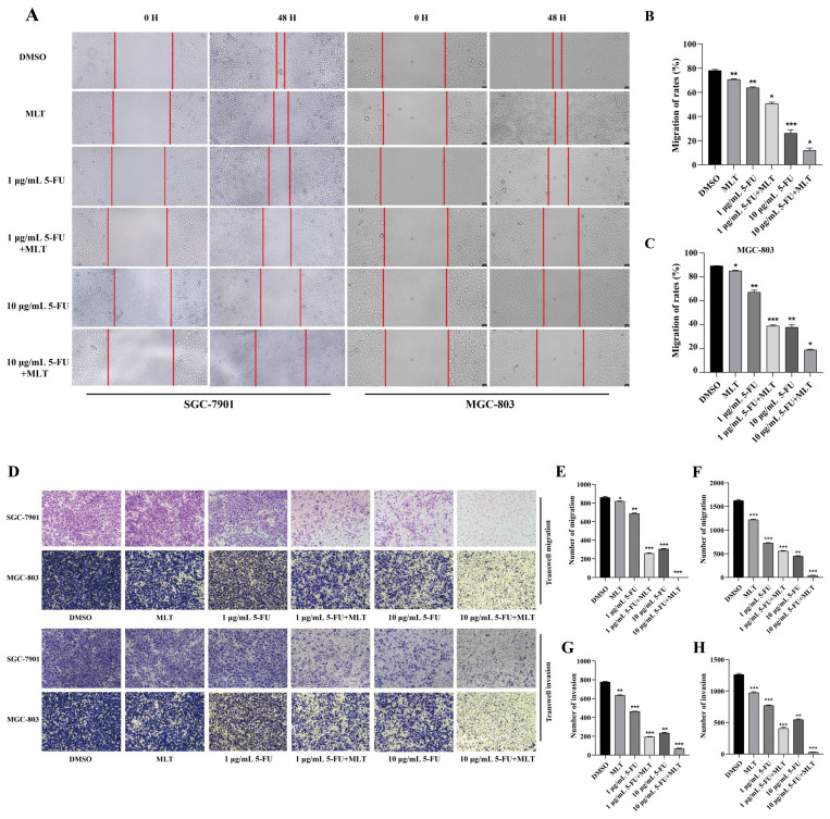
Figure 2.
Assessment of cell migration and invasion using wound-healing assay. (A) Gastric cancer (GC) cells were cultured to confluence. And the cell monolayer, thus formed, was damaged with a sterile tip of a 100 μL pipette. The cells were further treated with 5-fluorouracil (5-FU; 1 or 10 μg/mL) alone or in combination with melatonin (MLT; 1 mM). (B, C) The cells in culture were photographed and the width of the wound was measured at 0 and 48 h after treatment to calculate the percentage of cell migration. (D) Results of the Transwell migration experiment. (E-H) Invasion of SGC-7901 and MGC-803 cells treated with MLT or 5-FU alone or in combination for 48 h. The cells were photographed. And the percentage of invasive cells was calculated. The experiments were performed three times to ensure accuracy and consistency. One-way ANOVA with Tukey's multiple comparison test was used for statistical analysis. Data are presented as mean ± SD. And statistical significance is indicated using asterisks (*p < 0.05, **p < 0.01, ***p < 0.001).

