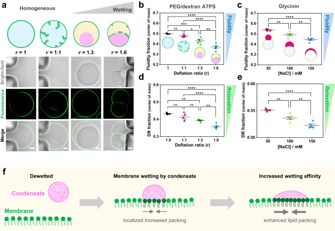
Fig. 8. Tuning membrane lipid packing and hydration is a general mechanism underlying wetting by condensate droplets. a.
DOPC vesicles labeled with 0.5 mol% LAURDAN filled with a PEG/dextran solution (ATPS) undergo morphological transformations due to the increase in membrane wetting affinity by the polymer-rich phases. Vesicle deflation with associated increases in the internal polymer concentration result from exposure to solutions of higher osmolarity. This leads to tube formation and subsequent phase separation in the vesicle (see Methods for further details). The dextran-rich phase (pink color in the sketch) is denser and has a higher refractive index105 than the PEG-rich phase (yellow), as can be observed in the bright-field images. The degree of vesicle osmotic deflation,, is given by the ratio of the external to initial internal osmolarity. The lower panel shows snapshots vesicles trapped in a microfluidic device subjected to different deflation ratios. The dark regions visible in the bright-field images are the microfluidic posts. Scale bars: 5 µm. b–e FLIM phasor fluidity and dipolar relaxation analysis for DOPC GUVs labeled with 0.5% LAURDAN in contact with the ATPS (b, d) and the glycinin condensates (c, e), respectively. The center of mass for fluidity changes (b, c) and dipolar relaxation (DR) changes (d, e) are shown. Histograms can be found in Supplementary Fig. 11. In both condensate systems, an increase in membrane wetting leads to a decrease in fluidity. Independent experiments are shown as circles and the lines represent mean values ± SD. The statistical analysis was performed with One-way ANOVA and Tukey post-test analysis (p < 0.0001, **** | p < 0.001, *** | p < 0.01, ** | p < 0.05, * | ns = non-significant). f Sketch summarizing the findings: when the condensate (pink) interacts with the membrane (green), wetting promotes an increased lipid packing (and decreased water dipolar relaxation) in the contact region. If the wetting affinity is increased, the effect on membrane packing becomes stronger. Note that the lipids in the contact region are colored in a darker green only for contrast, but the composition of the membrane remains unaltered. Data for panels (b-e) are provided as a Source Data file.

