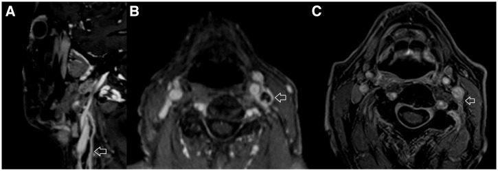
Figure 2.
MRI imaging demonstrating vagus nerve neuritis; brain imaging was normal. (A) Fat saturated T1 weighted post-contrast sagittal MRI of neck demonstrating enlarged left vagus nerve with avid peripheral enhancement (arrowed). (B) Fat-saturated T1 weighted post-contrast axial MRI of neck demonstrating enlarged left vagus nerve with avid peripheral enhancement (arrowed). (C) Fat saturated T1 weighted post-contrast axial MRI of neck, performed 6 weeks later, demonstrating almost complete resolution of the left vagus nerve changes. The arrow points to residual enhancement in the left carotid space in the region of the left vagus nerve.

