Fig. 5.
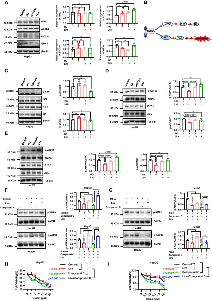
Liraglutide inhibits ferroptosis by activating AMPK/ACC signaling. A Western blotting show TFRC, ACSL4, SLC7A11 and GPX4 levels in HepG2 cells after 48-h incubation with 125 mM HG and 2.0 μM liraglutide. B Simplified schematic representation of the downstream pathway of AMPK. C Western blotting show phosphorylated S6K and phosphorylated S6 levels in HepG2 cells after 48-h incubation with 125 mM HG and 2.0 μM liraglutide. D Western blotting show phosphorylated AMPK levels and phosphorylated ACC levels in HepG2 cells after a 4-h incubation in glucose-free medium and a 4-h incubation with 125 mM HG, 2.0 μM liraglutide and combination. E Western blotting show phosphorylated AMPK levels and phosphorylated ACC levels in Hep3B cells after a 4-h incubation in glucose-free medium and a 4-h incubation with 125 mM HG, 2.0 μM liraglutide and combination. F Western blotting showing the levels of ACC (S79) and AMPK (T172) phosphorylation in HepG2 and Hep3B cells treated with 2.0 μM erastin for 12 h, or 5.0 μM Compound C for 12 h, 2.0 μM liraglutide for 48 h and the combination. G Western blotting showing the levels of ACC (S79) and AMPK (T172) phosphorylation in HepG2 and Hep3B cells treated with 1.0 μM RSL3 for 12 h, or 5.0 μM Compound C for 12 h, 2 μM liraglutide for 48 h and the combination. H Cell viability in HepG2 were measured treated as F. I Cell viability in HepG2 were measured treated as G. Data are expressed as mean ± SD, n = 4

