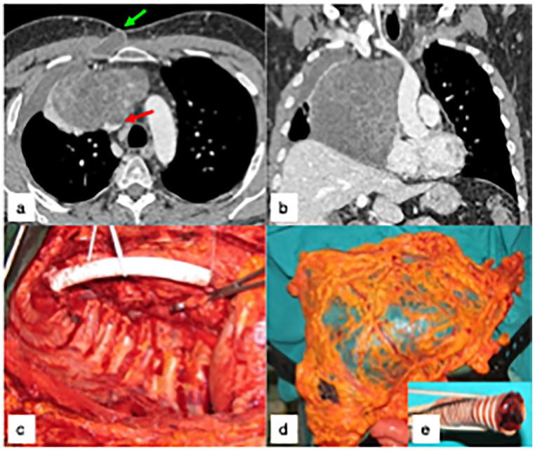
Figure 2.
(a-b) shows the preoperative CT of a 35 years-old male with primary mediastinal germ cell neoplasm invading the superior vena cava (SVC, red arrow), previously treated in another hospital by two cycles of PEB chemotherapy and 36Gy of external RT, with normalization of blood markers and apparent tumor size increase, presenting with a severe septic clinical picture, pleural empyema and cutaneous fistula (green arrow). The patient was treated by right extra-pleural pneumonectomy and SVC replacement with a PTFE prosthesis, through a thoraco-sternotomy approach (right hemi-clamshell, Figure 2c). Six months later, he was re-admitted for recurrent right pleural empyema with broncho-pleural fistula, and treated by a pedicled omental flap (Figure 2d) after removal of the infected PFE prosthesis (Figure 2e). The patient is alive and well, seventeen years later.

