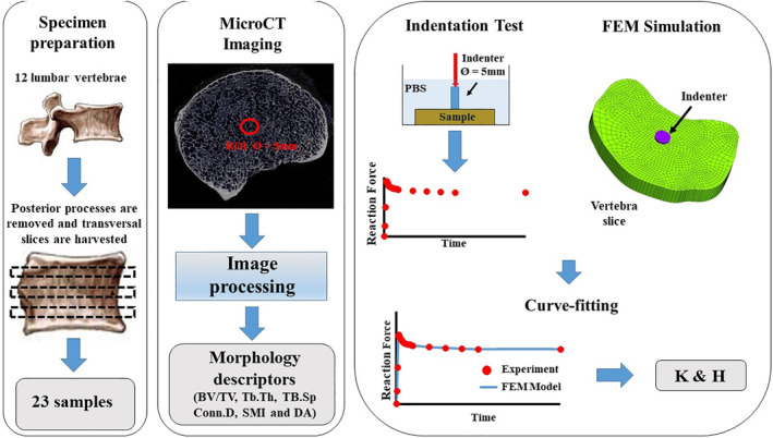
Figure 1.

Schematic of the workflow for this study. Twenty‐three transversal slices of vertebral bodies were obtained from 12 lumbar vertebras. μCT scanning provided bone morphology descriptors. Curve‐fitting of indentation tests with results from Finite Element Method (FEM) simulations yielded values of K and E. BV/TV = bone volume over total volume; Conn.D = connectivity density; DA = degree of anisotropy; SMI = structure model index; Tb.Sp = trabecular spacing; Tb.Th = trabecular thickness.
