
FIGURE 1.
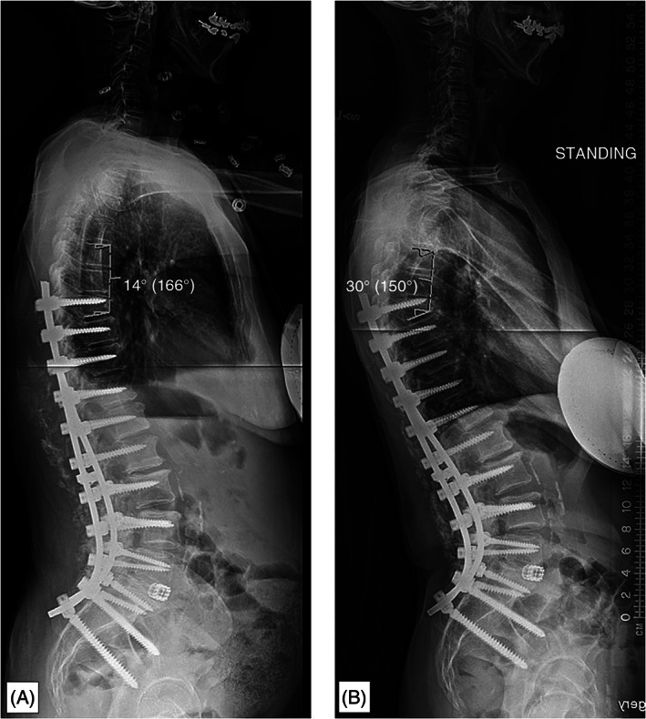
Radiographs illustrating (A) the technique used for measurement of proximal junctional angle (PJA) as well as (B) the same patient who went on to develop a 16° progression of the PJA, therefore meeting criteria as having developed proximal junctional kyphosis (PJK). 5