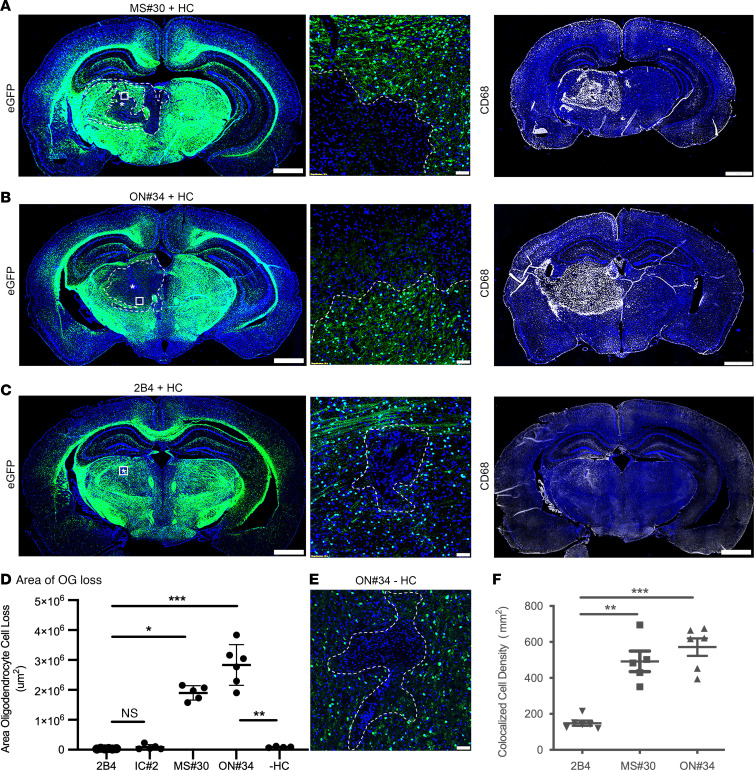
Figure 1. Myelin-specific rAbs initiate complement-dependent oligodendrocyte cell death.
(A–C) EGFP immunofluorescence in brain sections of C57BL/6 PLP-EGFP mice following ICI of myelin-specific (MS#30, ON#34) or IC (2B4) rAbs with HC. (Left panels) Amorphous regions of EGFP+ oligodendrocyte loss at 72 hours after injection are demarcated by the dotted lines. Asterisks indicate injection site. (Center panels) Higher magnification images of boxed areas reveal a sharp demarcation between areas of complete oligodendrocyte loss and adjacent normal-appearing tissue. (Right panels) CD68+ microglia/macrophages accumulate within the lesion core. Scale bars: 1 mm (left and right); 50 µm (center). (D) Quantitation of the area of EGFP+ oligodendrocyte cell loss (4–6 animals per injection) for IC (2B4, IC#2) rAbs plus HC, myelin-specific (MS#30, ON#34) rAbs plus HC, and myelin-specific ON#34 rAb minus HC (–HC) (Kruskal-Wallis 1-way ANOVA with Dunn’s correction for multiple comparisons, *P < 0.05; ***P < 0.001; ON#34 +/– HC, Mann-Whitney U test, **P < 0.01). (E) High-magnification image of ON#34 rAb without HC (ON#34 – HC) ICI shows a minor loss of EGFP+ oligodendrocyte cell loss at the injection site. Scale bar: 50 µm. (F) Quantitation of CD68+DAPI+ cell density (per subcortical hemisphere) at 72 hours after injection following ICI of rAbs 2B4, MS#30, or ON#34, plus HC (ANOVA with Dunn’s correction for multiple comparisons, **P < 0.01).

