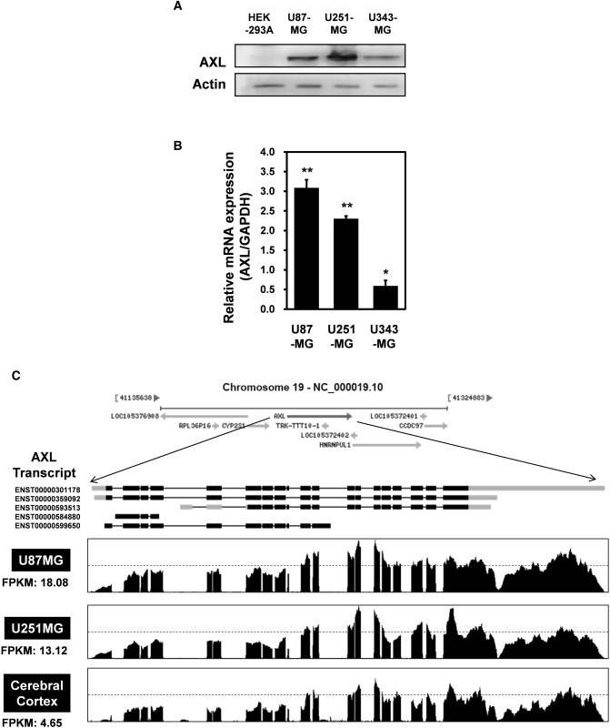
Fig. 1.
AXL expression in glioblastoma (GBM) cell lines. a Lysates were prepared from four established GBM cell lines (U87-MG, U251-MG and U373-MG) and subjected to western blotting using anti-AXL and anti-actin antibodies. The results are representative of three independent experiments (top panel). Relative densities were obtained by densitometry. Relative differences in AXL expression levels were calculated by normalizing all densitometric values to that of actin (in each lane). Results are presented as the means ± SDs of data from three independent experiments. b Total RNA extracted from each GBM cell line was analyzed by real-time quantitative reverse transcription-polymerase chain reaction (qRT-PCR) using human AXL-specific primers, as described in Materials and Methods. C Total RNAs were isolated from two GBM cell lines (U87-MG and U251-MG) and normal brain tissue. These samples were analyzed by standard RNA deep-sequencing (RNA-seq), as described in Materials and Methods. RNA-seq read densities of AXL transcripts were plotted against relative RNA-seq read coverages (counts). “Fragments per kilobase of exon per million fragments mapped” (FPKMs) were calculated to compare the expression levels of AXL mRNA variants among various samples. The results are presented as means ± SDs of data from three independent experiments. *p < 0.05, **p < 0.01

