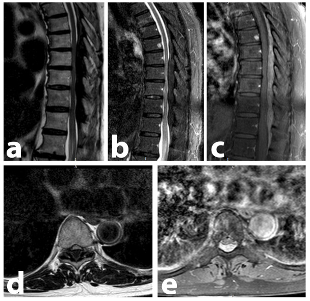
Figure 1.
: Thoracolumbar spine MRI performed at patient admission, demonstrating an extensive posterior extradural lesion, ranging from T6 to L1, causing spinal cord compression. Compression is most expressive between the T8-T11 levels. There are no bone lesions consistent with MM. (A) Sagittal T1-weighted image showing the hypointense lesion. (B) Sagittal STIR image showing the hypointense lesion. (C) Sagittal contrast-enhanced T1 SPIR image, where the lesion demonstrates a diffuse and homogeneous enhancement to the paramagnetic contrast medium. This is the image that most clearly demonstrates the full extent of the lesion. (D and E) These T9-level axial images, T1-weighted and T1-enhanced SPIR, demonstrate the extrinsic compression caused by the posterior epidural mass (hypo and hyperintense, respectively).

