
Figure 3.
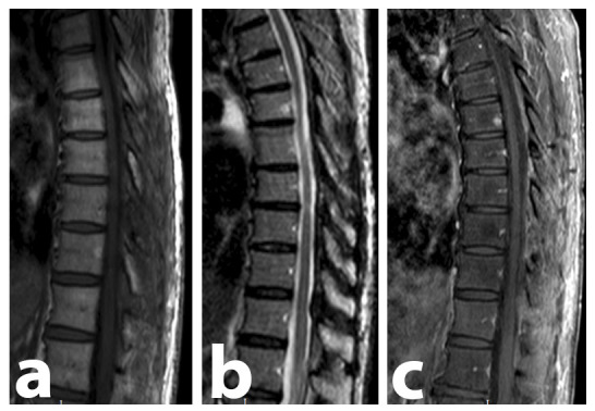
Thoracolumbar spine MRI performed 8 months after surgery. No lesion remains could be found, proving the complete resection of the lesion, as well as the absence of recurrence. There are no bone lesions consistent with MM. (A) Sagittal T1-weighted image. (B) Sagittal STIR-weighted image. (C) Sagittal contrast-enhanced T1-weighted SPIR image demonstrating the absence of contrast-enhancing lesions.