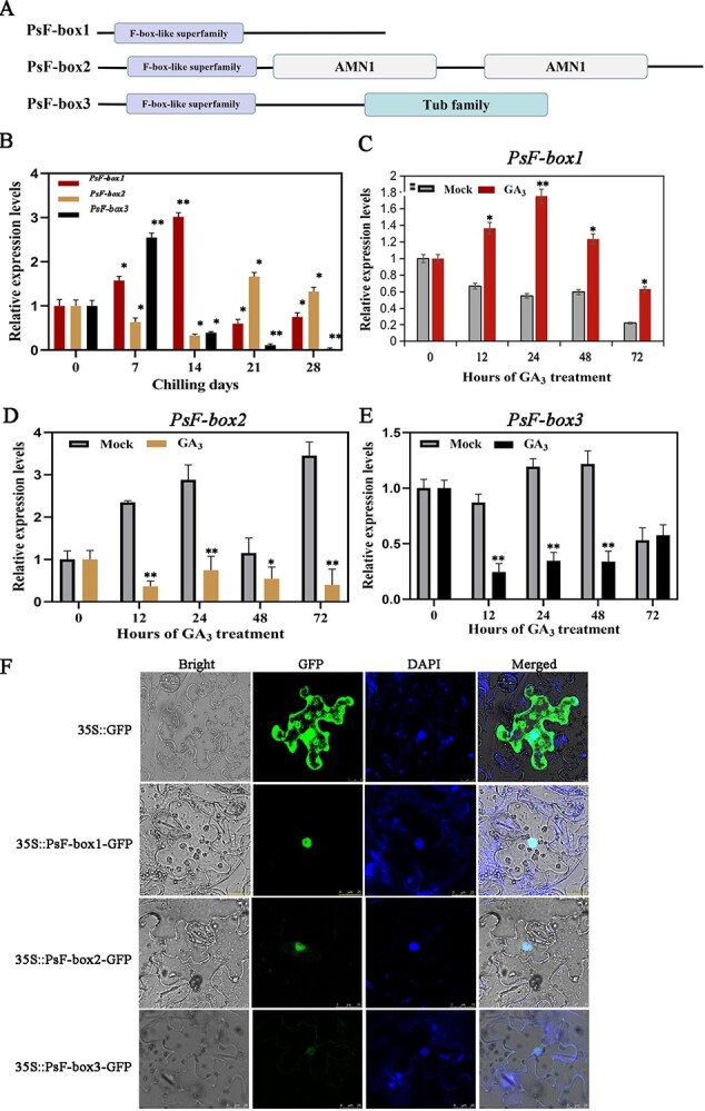
Figure 2.

Domain arrangement of the putative PsF-box proteins and the expression patterns of three PsF-box genes during chilling- and GA3-induced dormancy release. A Linear representation of the domain arrangement of the putative PsF-box proteins, of which PsF-box1 had a 567 bp ORF encoding 189 aa, PsF-box2 had a 1617 bp ORF encoding 539 aa, and PsF-box3 had a 777 bp ORF encoding 259 aa. Boxes with different colors represent different domains. B Expression patterns of PsF-box1, PsF-box2, and PsF-box3 after different chilling durations. C–E Expression of PsF-box1, PsF-box2, and PsF-box3 after exogenous GA3 treatment, respectively. F Subcellular localization of PsF-box1, PsF-box2, and PsF-box3 by fluorescence microscope with an excitation wavelength of 488 nm. Data represent the mean ± SD of six replicates. *P < 0.05; **P < 0.01.
