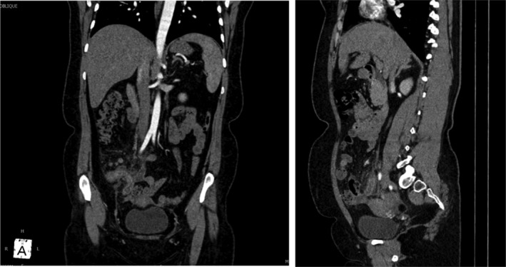
FIGURE 1.

Contrast‐enhanced computed tomography of abdomen and pelvis (a‐ coronal and b‐ saggital) showing enlarged appendix with fat strandings in the periappendiceal region, and mucosal enhancement in the terminal ileum, ileocecal valve, and nearby cecum.
