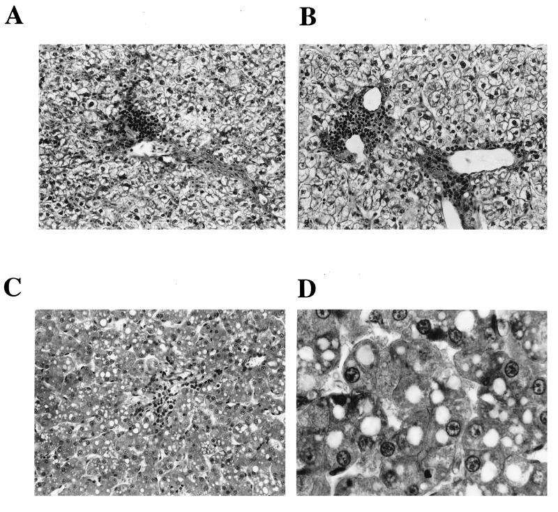
FIG. 4.
Analysis of liver histology revealing microvesicular steatosis in ddC-treated animals and no histological sign of toxicity during short-term administration of l-FMAU. (A) Liver histology for one control animal. Signs of viral hepatitis are observed: ballooning hepatocytes, portal tract infiltration, and lobular inflammation (obj., 25). (B) Liver histology for one l-FMAU (40 mg/kg every day)-treated animal. A pattern similar to that in the control animals is observed. (C) Liver histology for one ddC (50 mg/kg twice a day)-treated duckling. Typical signs of microvesicular steatosis (accumulation of lipid droplets in the cytoplasms of hepatocytes) and acidophilic necrosis of hepatocytes are shown. (D) Liver histology for the same ddC (50 mg/kg twice a day)-treated duckling but at a higher magnification (obj., 40), which shows intracytoplasmic lipid droplets.

