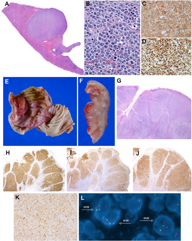
Fig. 4.
Histologic and immunophenotypic features of large B-cell lymphoma with IRF4-rearrangement presenting in extranodal sites. A–D Case LYWS-1163 courtesy of E. Shuyu. A Spleen section showing a well-circumscribed nodular infiltration. B The tumor is composed of large centroblastic lymphoid cells with open chromatin, several large nuclei, and abundant cytoplasm. C The tumor cells are CD10 and D IRF4/MUM1 strongly positive. E–L Case LYWS-1112 courtesy of D. Wang. E Terminal ileum with a 2.2-cm large polypoid mass. F The intestinal crossed section shows a white soft mass infiltrating the mucosa, submucosa, and the muscularis propria. G H&E section reveals a follicular lymphoid infiltrate with large, back-to-back follicles. The follicles are composed of medium to large-sized centroblasts. H The tumor cells are positive for CD79a, I CD10, J BCL6, and K IRF4/MUM1. L Interphase FISH analysis using break apart probes for IRF4. Most cells have 3 fusion signals (yellow) and 2 red signals with loss of the green signals indicating an IRF4 translocation

