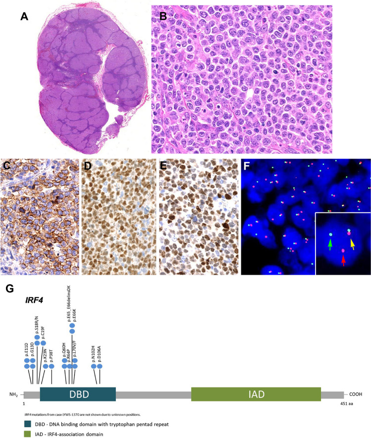
Fig. 6.
Histologic and immunophenotypic features of large B-cell lymphoma with IRF4-rearrangement in an adult patient. Case LYWS-1370 courtesy of B. Bisig. A Panoramic view of a lymph node with preserved capsule and effacement of the architecture by the presence of multiple, large confluent nodules. No diffuse areas or necrosis are observed. B Higher magnification demonstrates that the nodules are composed of large cells with fine chromatin, multiple nucleolei, abundant cytoplasm typical of centroblasts. Some apoptotic figures and some mitosis are observed. C The tumor cells were CD10 positive as well as D BCL6 and E IRF4/MUM1. F Interphase FISH analysis using break apart probes for IRF4 revealed 1 fusion signal (yellow arrow), one red signal (red arrow), and one green signal (green arrow) indicative of an IRF4 rearrangement. G Diagram of the relative positions of IRF4 driver mutations. The approximate location of somatic mutations identified is indicated. The analysis was performed by next generation sequencing. IRF4 mutations are mainly in the DNA binding domain (DBD). Domains of the protein are represented according to the Uniprot database (www.uniprot.org)

