Fig. 7.
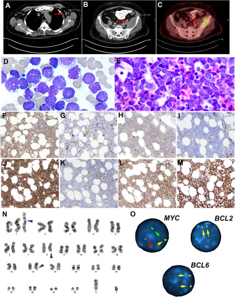
Histologic, immunophenotype, and cytogenetic features of a B-cell acute lymphoblastic leukemia with IGH::MYC according to the 2022 ICC. Case LYWS-1026 courtesy of G. Caponetti. A CT scan demonstrates supradiaphragmatic lymphadenopathy (red arrow). B CT scan reveals a soft tissue lesion expanding the posterior aspect of the left iliacus muscle (red arrow) C PET scan shows intense FDG uptake by the soft tissue mass (red arrow). D The bone marrow aspirate shows 81% blasts with vacuolated cytoplasm typical of Burkitt lymphoma. E The bone marrow biopsy is hypercellular for age and shows a diffuse infiltrate with relatively large blastoid cells. F The tumor cells are positive for CD19. G CD10, H TdT, and I CD34 are negative. J CD10 is strongly positive in the tumor cells. K BCL2 is negative, whereas L MYC is strongly positive. M The MIB1 stain shows a 100% proliferation. N The karyotype analysis reveals an abnormal male karyotype with a supernumerary isochromosome for the entire long arm of chromosome 1, which results in tetrasomy 1q (black arrow head). Additionally, a balanced translocation between chromosome 8 and 14 is observed with breakpoints at bands 8q24.2 and 14q32 resulting in a IGH::MYC translocation. O Interphase FISH analysis using break apart probes for MYC, BCL2, and BCL6 reveals in MYC 1 fusion signal (yellow arrow), one red signal (red arrow), and one green signal (green arrow) indicative of MYC rearrangement. In contrast, the analyses of BCL2 and BCL6 demonstrate two normal fusion signals (yellow arrows)

